LCGC Europe Application Note Alert
Having trouble viewing this email? Click here
Spectroscopy
Raman Imaging and Correlative Microscopy Techniques
Raman imaging can be combined with topographic Raman imaging, fluorescence, and scanning electron microscopy. This paper gives examples of its correlative potential.
Download Application Note
Characterizing Polymer Blends with Correlative Microscopy
This study shows Raman imaging, atomic force microscopy, and scanning near-field optical microscopy characterizing and depth-profiling polymer blends and layers.
Download Application Note
Characterizing Pharmaceutical Samples with Raman Microscopy
Pharmaceutical research benefits from Raman microscopy, which characterizes distributions, homogeneity, drug states, and contaminants. This study provides examples.
Download Application Note
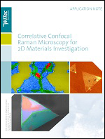
Correlative Raman Microscopy for 2D Materials
Two-dimensional (2D) materials such as graphene and transition metal dichalcogenides are well-suited to investigation by Raman and SEM. Examples of both are presented in the following app note.
Download Application Note
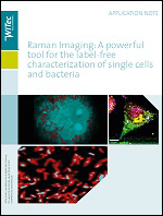
Raman Imaging: Single Cells and Bacteria
In this application note, 3D confocal Raman imaging is used to chemically characterize single cells and bacteria, nondestructively and without specialized preparation.
Download Application Note
Diagnostics and Treatment with Raman Imaging
Biological and medical research benefits greatly from Raman imaging and correlative microscopy. This note shows applications that demonstrate its advantages.
Download Application Note
 
 
UBM