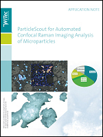
This overview of advanced microparticle analysis shows how particles can be found, classified and then identified automatically using confocal Raman microscopy.
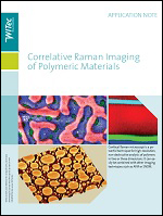
This study shows Raman imaging, atomic force microscopy and scanning near-field optical microscopy characterizing and depth-profiling polymer blends and layers.
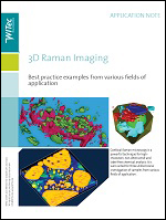
This study describes the generation of 3D images of pharmaceutical, biological and geological samples using image stacking enabled by a highly confocal beam path.
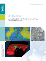
This paper details RISE (Raman Imaging and Scanning Electron) microscopy and demonstrates its advantages using geological, polymer, biological and nanomaterial samples.
In this application note, 3D confocal Raman imaging is used to chemically characterize single cells and bacteria, non-destructively and without specialized preparation.
2D materials such as graphene and transistion metal dichalcogenides are well-suited to investigation by Raman and SEM. Examples of both are presented in the following.