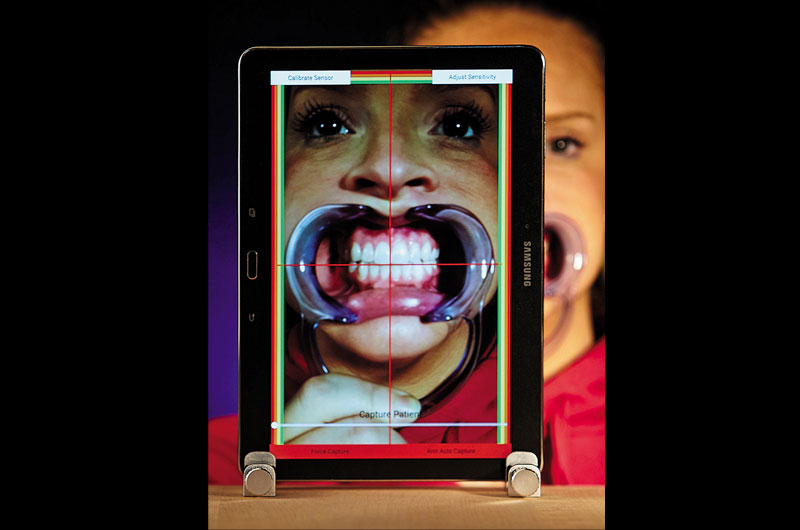
Fig. 01 Taking the photograph of the patient.
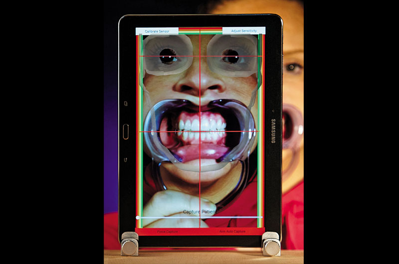
Fig. 02 Properly measuring the image.
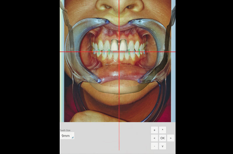
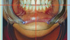
Fig. 03 Determining the first point of contact and the width of the centrals.
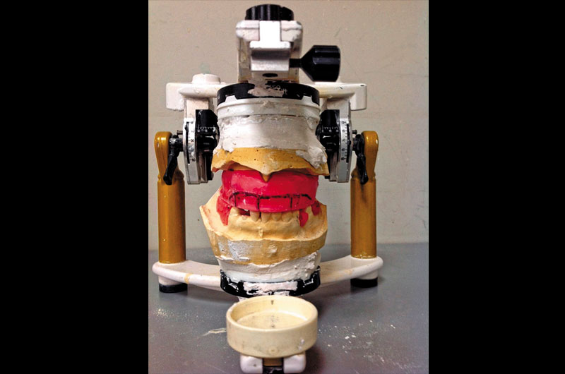
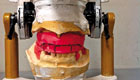
Fig. 04 A mounted case that looks right—but isn’t.
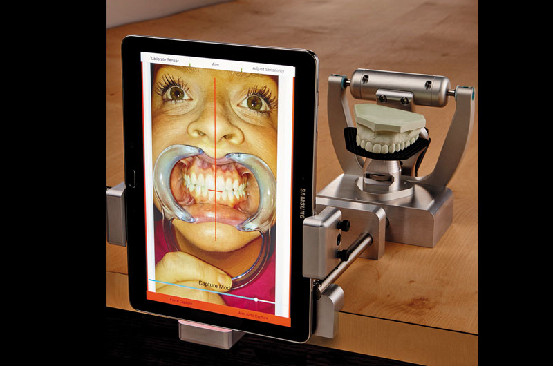
Fig. 05 Using MaxAlign in the lab with a stand.
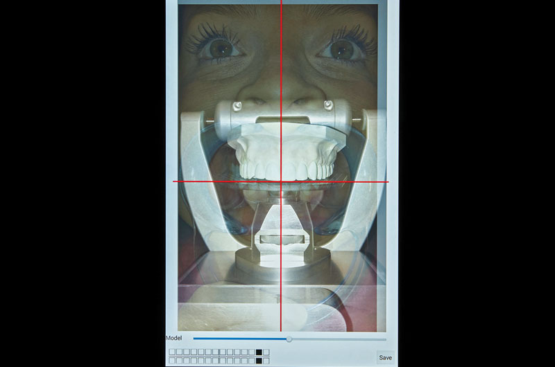
Fig. 06 The lab uses a model for verifications via the MaxAlign.