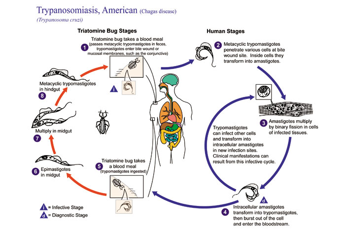
Fig. 1 This illustration depicts the life cycle of Trypanosoma cruzi, the causal agent of American Trypanosomiasis.

Fig. 2 This is a micrograph of Trypanosoma cruzi in a blood smear using Giemsa staining technique.
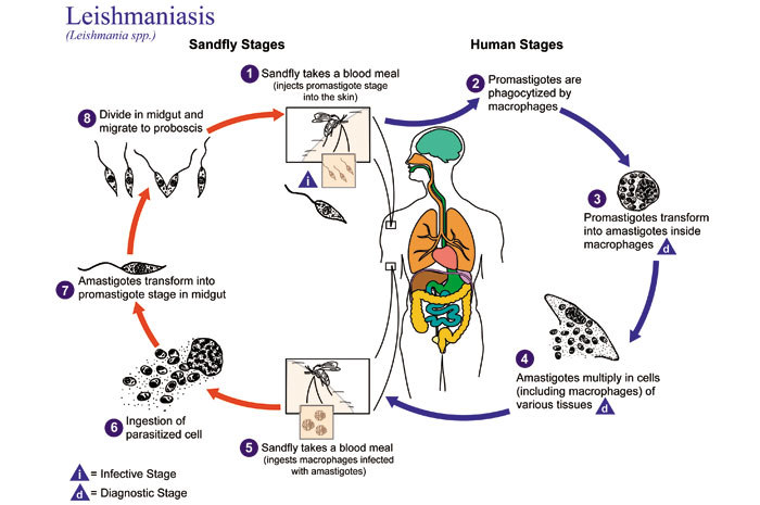
Fig. 3 This is an illustration of the life cycle of Leishmania spp., the causal agents of Leishmaniasis.
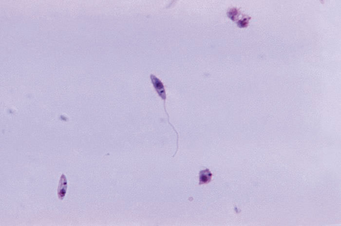
Fig. 4 This micrograph revealed the presence of a number of leptomonad-staged, i.e. promastigote, Leishmania sp. protozoa.

Fig. 5 Histopathology of leishmaniasis of skin due to Leishmania brasiliensis. Parasite.
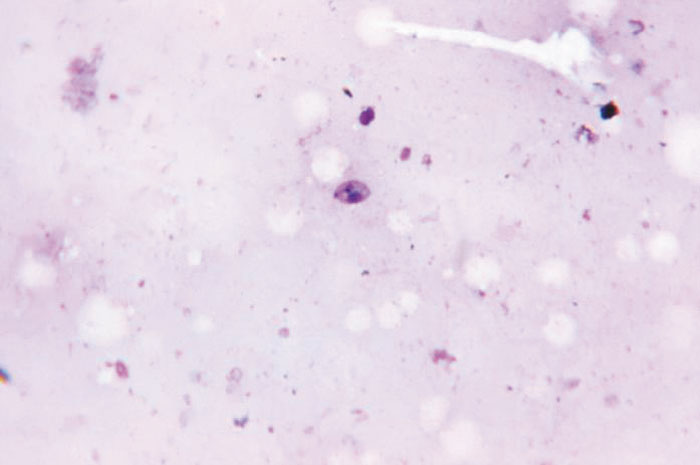
Fig. 6 This Giemsa-stained photomicrograph revealed a Leishmania sp. protozoan, which had been taken from a smear of a suspected leishmanial lesion, and magnified 1125X.
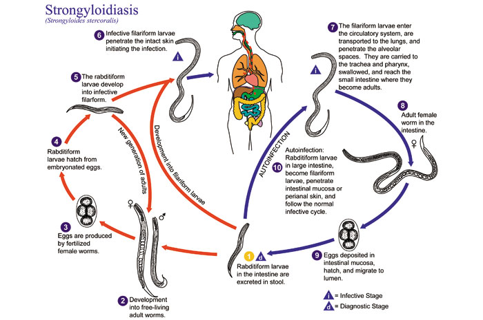
Fig. 7 This is an illustration of the life cycle of Strongyloides stercoralis, the causal agent of Strongyloidiasis.
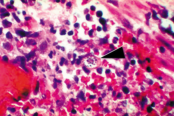
Fig. 8 This micrograph reveals a Strongyloides stercoralis parasite embedded in the intestinal mucosa.
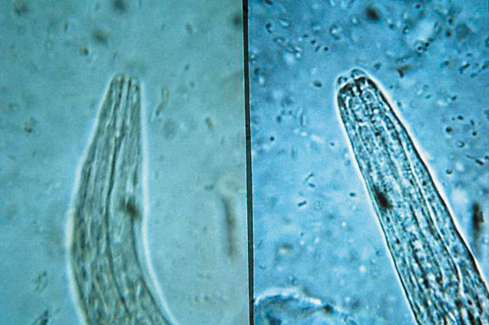
Fig. 9 This micrograph depicts the mouth parts of rhabditiform staged larvae of a hookworm (Lt), and strongyloides (Rt); Mag. 500X.
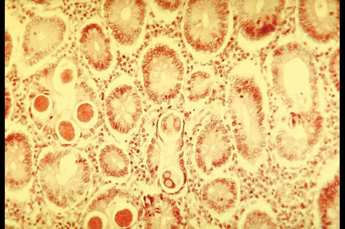
Fig. 10 This photomicrograph depicted numbers of Strongyloides parasites found inside an intestinal tissue specimen.
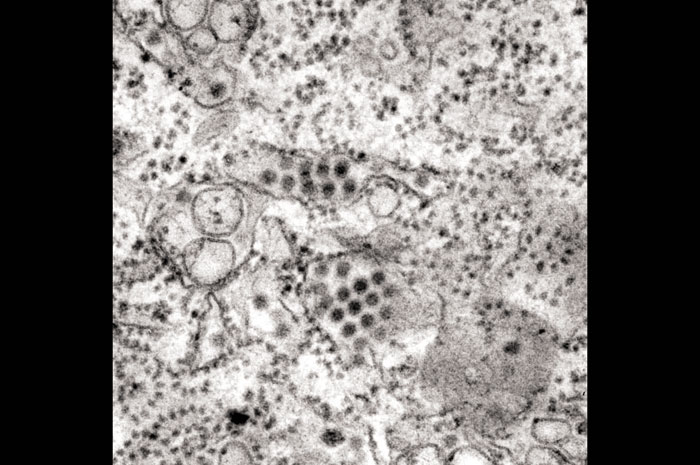
Fig. 11 This transmission electron micrograph (TEM) depicts a number of round, Dengue virus particles that were revealed in this tissue specimen.
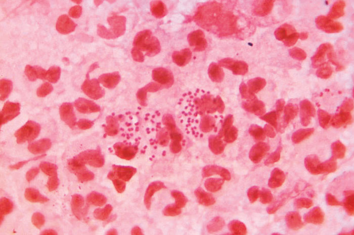
Fig. 12 This Gram-stained photomicrograph reveals the presence of intracellular Gram-negative, Neisseria gonorrhoeae diplococcal bacteria, amongst numerous white blood cells (WBCs) known as polymorphonuclear leukocytes, or PMNs.
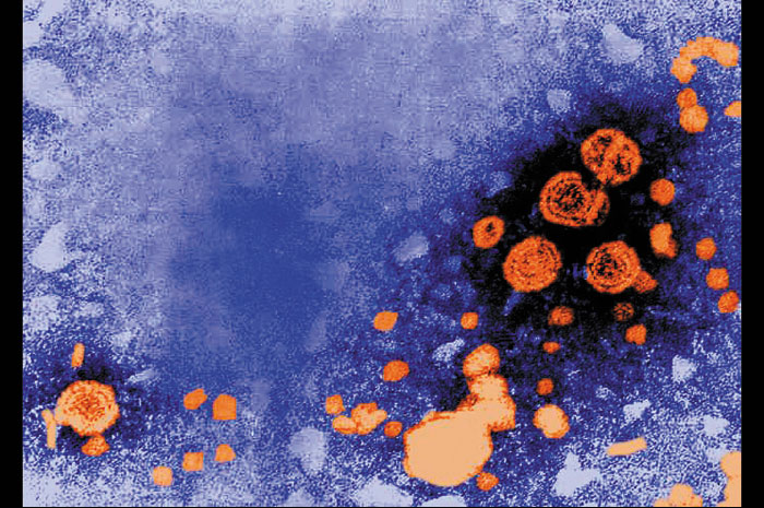
Fig. 13 This digitally-colorized transmission electron micrograph (TEM) revealed the presence of hepatitis B virions. The large round virions are known as Dane particles. See PHIL 270, for a black and white version of this image.
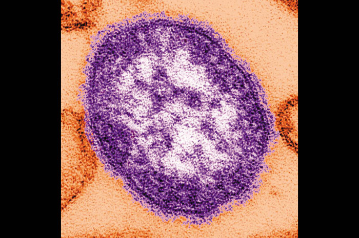
Fig. 14 This thin-section transmission electron micrograph (TEM) revealed the ultrastructural appearance of a single virus particle, or “virion”, of measles virus. The measles virus is a paramyxovirus, of the genus Morbillivirus. It is 100-200 nm in diameter, with a core of single-stranded RNA, and is closely related to the Rinderpest and canine distemper viruses. Two membrane envelope proteins are important in pathogenesis. They are the F (fusion) protein, which is responsible for fusion of virus and host cell membranes, viral penetration, and hemolysis, and the H (hemagglutinin) protein, which is responsible for adsorption of virus to cells.
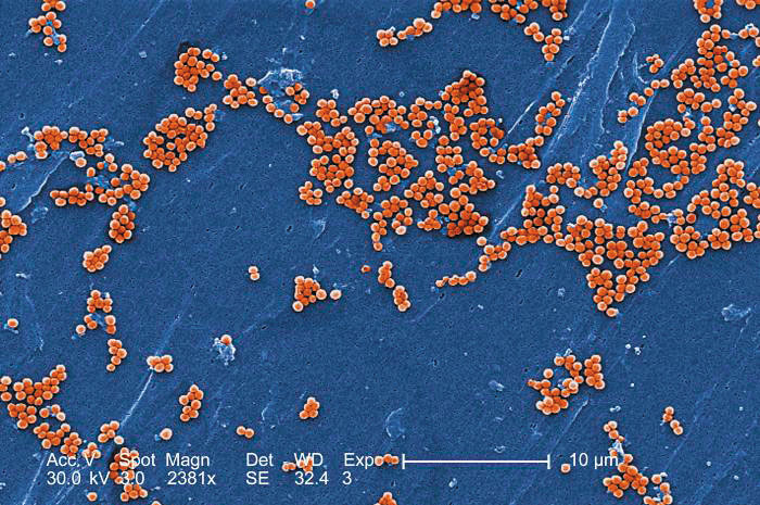
Fig. 15 This 2005 scanning electron micrograph (SEM) depicted numerous clumps of methicillin-resistant Staphylococcus aureus bacteria, commonly referred to by the acronym, MRSA; Magnified 2381x.
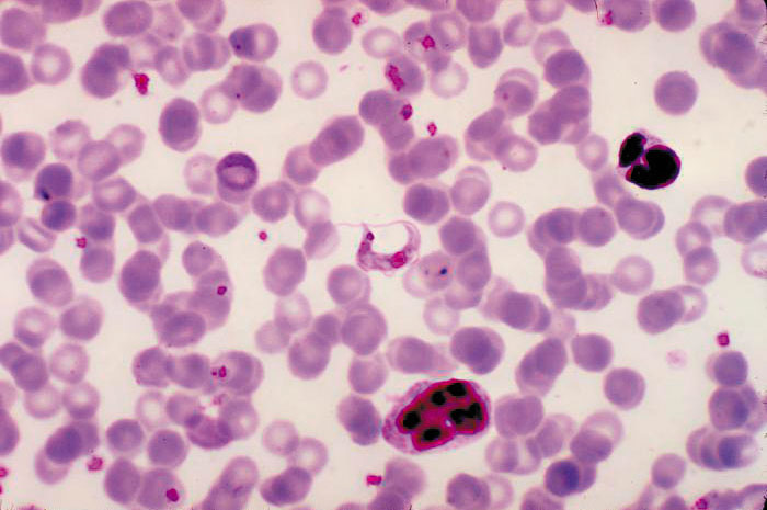
Fig. 16 Under a high magnification of 10,000x, this scanning electron micrograph (SEM) shows a strain of Staphylococcus aureus bacteria taken from a vancomycin intermediate resistant culture (VISA).
Fig 1
Fig 2
Fig 3
Fig 4
Fig 5
Fig 6
Fig 7
Fig 8
Fig 9
Fig 10
Fig 11
Fig 12
Fig 13
Fig 14
Fig 15
Fig 16