A protein misfold represents a structural impurity and at any level can result in changed efficacy and increase the potential for immunogenicity. Measuring low level impurities in secondary structure is necessary to maintain confidence in a protein’s integrity during all phases of drug development. FTIR and Circular Dichroism (CD) are commonly used for this, but have known limitations in reproducibility
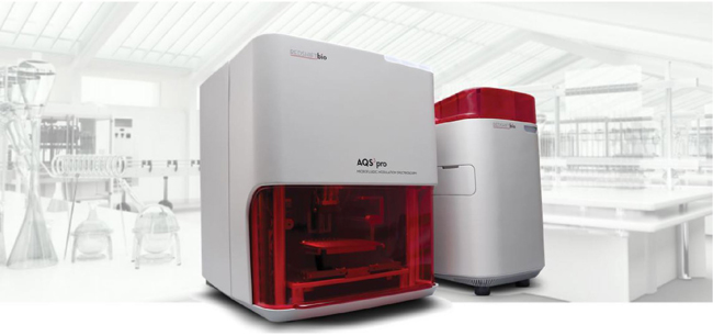
and sensitivity. Microfluidic Modulation Spectroscopy (MMS) is a new protein characterization method
which generates reproducible high resolution measurements, directly addressing the shortcomings of FTIR and CD.
This whitepaper from RedShiftBio directly compares the three techniques.
The lab used BSA spiked into IgG1 as this mimics a ‘best-case’ ability to detect a very different structural impurity (IgG1 is mainly β-sheet, while BSA is mainly α-helix). This whitepaper demonstrates that MMS out-performs traditional FTIR and CD methods across a number of parameters.